
|
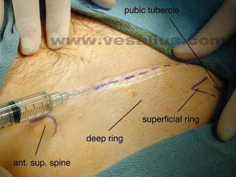
| This image shows the right groin. The inguinal canal was located using bony landmarks. |
|
| The incision is made down to the level of the superficial ring. The needle is injecting anesthetic to the area. |
|
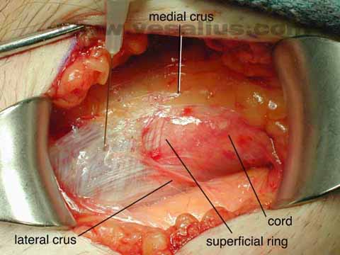
| The ilioinguinal nerve has been dissected out and is being preserved by placing it lateral to a flap of the external oblique aponeurosis. |
|
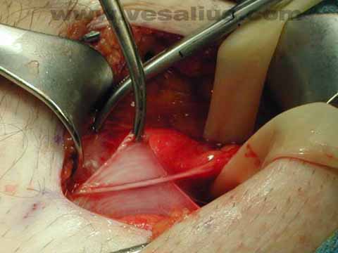
| The bulge of the direct hernia becomes clear. |
|
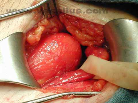
| A polypropylene mesh and absorbable sutures are used to repair the defect. |