|
|
|
||||||||||||
Dissector Answers - Joints of the Upper Limbs |
|||||||||||||
Learning Objectives:
Upon completion of this session, the student will be able to:
- List and describe the various types of moveable joints and give examples of each type.
- List the characteristics of and identify the parts of a typical synovial joint.
- Recall the movement characteristics of the various types of synovial joints.
- Identify the bony, cartilagenous, ligamentous and membranous components of the following joints:
- List the movements permitted at each joint and the ligaments that restrict them.
- Correlate joint movements with the muscles producing these actions at each joint.
Learning Objectives and Explanations:
1. List and describe the various types of moveable joints and give examples of each type. (W&B 46-51)The three major types of joints, along with subtypes and examples, are listed here:2. List the characteristics of and identify the parts of a typical synovial joint. (W&B 47-49)
- Fibrous joints: the most simple joints. They are only connected by fibrous ligaments. A suture is a fibrous joint that eventually fuses, forming one bone from two (a synostosis), like in the skull of a growing infant. A gomphosis is the joint between a tooth and the jaw. A syndesmosis is a fibrous membrane or ligament that joins two bones. The tibia and fibula have an interosseous ligament or membrane, as do the radius and ulna.
- Cartilaginous joints: joined by cartilage only. These joints are avascular or anervous, except at their margins. Synchondroses are temporary joints present in growing bones. The epiphyseal plate (growth plate) will later ossify into solid bone. The epiphysis has hyaline cartilage and the extension of ossification from the diaphysis side converts it to bone. A symphysis is a permanent cartilaginous union. They always have hyaline cartilage on the bony surfaces concerned, and these cartilaginous surfaces are joined by fibrous tissue or fibrocartilage.
- Synovial joints: "diarthroses" (freely moveable joints). These joints are joined by a fluid-filled capsule and accessory ligaments. Examples include the knee, ankle, and hip.
Synovial joints consist of:3. Recall the movement characteristics of the various types of synovial joints. (W&B 47-49)Types of synovial joints
- Hyaline cartilage: covers the full weight-bearing surface, providing a smooth yet resilient surface
- Joint capsule: a cavity, made of accessory ligaments, with synovial fluid inside. This reinforces the synovial membrane.
- Synovial membrane lining: secretes synovial fluid and covers the synovial cavity. It reaches to the edges of the hyaline cartilage.
- Accessory structures: accessory ligaments ("intracapsular" and "extracapsular"); articular discs or menisci, which are pads of fibrous cartilage; muscles and tendons; and subsynovial fat.
- Plane: involves flat surfaces. Movements consist of sliding of one surface on the other, and may be multidirectional in one plane. Examples: facet joints, joints of the tarsal bones of foot.
- Hinge (ginglymus): movement around a single axis at right angles to the bone. Permits flexion and extension only. These usually have strong collateral ligaments on each side reinforcing the joint. Examples: elbow, knee.
- Pivot (trochoid): rotary movement around a longitudinal axis. Rounded process of bone rotates within a sleeve or ring composed of a bony fossa and a strong ligamentous band. Examples: atlas-axis, radioulnar joint.
- Condyloid: oval surfaces allowing movements in two planes at right angles to each other. Example: radiocarpal joint
- Saddle (sellar): movement in two basic axes, with circumduction. Example: carpal-metacarpal joint of the thumb.
- Ball and socket: allows movement in any axis. Examples: hip, shoulder.
See #2 above.4. Identify the bony, cartilaginous, ligamentous and membranous components of the following joints (W&B 169-183):
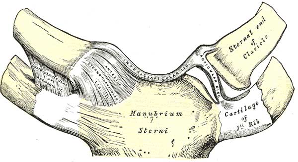
sternoclavicular joint: (TG2-42A, TG2-42BC)5. List the movements permitted at each joint and the ligaments that restrict them. (W&B 169-183)
The articular disc of the sternoclavicular joint serves to absorb shock as force is transmitted along the clavicle. Its joint capsule is formed from the sternoclavicular ligament, which is divided into anterior and posterior parts.
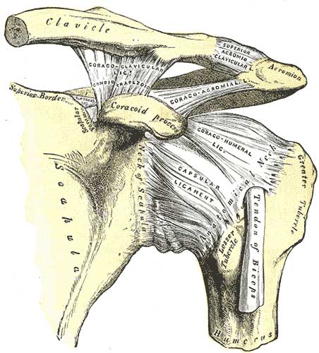
acromioclavicular and shoulder joints: (TG2-42A, TG2-42BC)
The important components of the acromioclavicular joint are the coracoacromial and the coracoclavicular ligaments. The former connects the coracoid process to the acromion, while the latter is a connection between the coracoid process and the clavicle. An injury to this joint is called a "shoulder separation". See #4 below.
The shoulder joint (glenohumeral joint) is the most mobile joint in the body. It has the following important components:
- glenohumeral bands: superior, middle, and inferior capsular ligaments that connect the humerus to the glenoid process
- subscapular bursa: protects the subscapular tendon where it passes inferior to the coracoid process and over the scapular neck
- tendon of long head of biceps: from its origin on the supraglenoid tubercle of the scapula, the tendon passes through the capsule of the shoulder joint into the intertubercular groove of the humerus, enclosed by a synovial sheath.
- transverse humeral ligament: spans the intertubercular groove, holding the tendon of the long head of the biceps in place
- glenoid labrum: much like the acetabular labrum, it deepens the socket and helps to hold the humerus in the glenoid fossa.
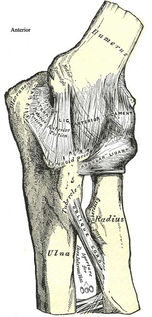
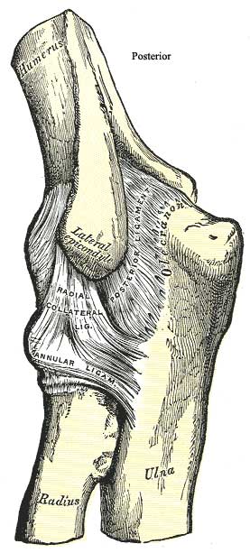
elbow (humeroulnar, humeroradial, proximal radioulnar) joint: (TG2-43A, TG2-43BC)
The most important structures here are the ulnar and radial collateral ligaments and the anular ligament. The radial collateral ligament is lateral, and extends from the lateral epicondyle of the humerus and blends distally with the anular ligament of the radius. The ulnar collateral ligament extends from the medial epicondyle of the humerus to the coronoid process and olecranon of the ulna. Finally, the anular ligament encircles the head of the radius. It holds the head of the radius against the ulna and provides restraint against distal dislocation of the radius
distal radioulnar joint: (TG2-44A, TG2-44B, TG2-44C)
This joint allows the distal end of the radius and the distal end of the ulna to rotate about one another, which is necessary for pronation and supination. It has an intracapsular articular disc.
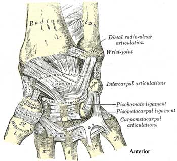
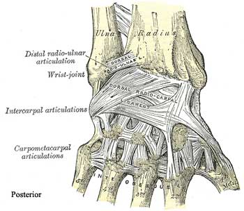
wrist (radiocarpal) joint and joints of the hand: (TG2-44A, TG2-44B, TG2-44C)
This joint is comprised of two radiocarpal ligaments. The dorsal radiocarpal ligament allows the hand to follow the radius during pronation of the forearm, while the palmar radiocarpal ligament allows the hand to follow the radius during supination. There are also two collateral ligaments, ulnar and radial, that prevent hyperabduction and hyperadduction respectively.
There are articulations between each row of bones in the hand, namely intercarpal, carpometacarpal, metacarpophalangeal (MP), and interphalangeal (IP) joints:
Joint Description Significance carpometacarpal joint, thumb synovial, saddle (concavoconvex) connects trapezium with metacarpal of thumb; flexion/extension, abduction/adduction carpometacarpal joints, fingers synovial, plane connects distal carpal bones with metacarpals of fingers; tightly bound by ligaments to limit motion metacarpophalangeal joints synovial, condyloid or ellipsoid connects metacarpal head to base of proximal phalanx; strengthened by collateral ligaments; heads of metacarpals are firmly joined by transverse metacarpal ligaments to provide a stable platform for finger movements interphalangeal joints synovial, hinge connect proximal & middle phalanges (proximal interphalangeal joint) and middle & distal phalanges (distal interphalangeal joint); strengthened by collateral ligaments 6. Correlate joint movements with the muscles producing these actions at each joint. (W&B 169-183)
- sternoclavicular joint: can move anteriorly, posteriorly, and inferiorly to allow movement of the clavicle along with movements of the upper limb. Movements are restricted by the sternoclavicular ligament.
- acromioclavicular joint: though there are no muscles that directly move the bones of this joint, there is some movement allowed as the whole upper limb is moved, though things are held in place by the coracoacromial and coracoclavicular ligaments.
- shoulder (glenohumeral) joint: almost anything is possible in the most mobile joint of the body. It can flex, extend, abduct, adduct, rotate medially, and rotate laterally. All of the ligaments described above, including the glenohumeral bands, the tendon of long head of biceps, the transverse humeral ligament, and the glenoid labrum help hold this joint together, albeit relatively loosely.
- elbow joint: this hinge joint can only flex and extend. Clinically, the most important ligament in terms of keeping the joint together is the anular ligament.
- proximal and distal radioulnar joints: allow pronation and supination of the forearm.
- wrist joint: capable of flexion, extension, adduction, and abduction. More flexion is possible than extension, and both are augmented by movements of the joints within the hand. There is more adduction possible than abduction, and the latter occurs mostly at the midcarpal joint, not the wrist joint.
- carpometacarpal joints: with the thumb and little finger these joints are quite mobile, allowing flexion, extension, adduction, and abduction. The joints of the middle three digits, however, are quite immobile.
- metacarpophalangeal (MP) joints: allow flexion, extension, adduction, and abduction.
- interphalangeal (IP) joints: allow only flexion and extension.
- sternoclavicular joint: no direct muscle action
- acromioclavicular joint: no direct muscle action
- shoulder (glenohumeral) joint:
- flexors: pectoralis major, deltoid (anterior fibers), coracobrachialis, biceps brachii
- extensors: latissimus dorsi, assisted by teres major
- abductor: deltoid (central fibers, following initiation by supraspinatus)
- adductors: pectoralis major and latissimus dorsi
- medial rotator: subscapularis, assisted by pect. major, latissimus, teres major
- lateral rotators: infraspinatus, teres minor
- elbow joint:
- flexors: brachialis, biceps brachii, brachioradialis, pronator teres (somewhat)
- extensors: triceps brachii, anconeus
- proximal and distal radioulnar joints:
- supinators: supinator, biceps brachii, extensor pollicis longus (somewhat), extensor carpi radialis longus (somewhat)
- pronators: pronator quadratus, pronator teres, flexor carpi radialis (somewhat), palmaris longus (somewhat), brachioradialis (somewhat)
- wrist joint:
- flexors: flexor carpi radialis, flexor carpi ulnaris, flexors of digits (somewhat), palmaris longus (somewhat), and abductor pollicis longus (somewhat)
- extensors: extensor carpi radialis longus and brevis, extensor carpi ulnaris, extensors of fingers and thumb (somewhat)
- carpometacarpal joints: intrinsic hand muscles and other muscles of the hand, especially relating to the thumb and little finger
- metacarpophalangeal (MP) joints: interosseous muscles, intrinsic and extrinsic abductors, flexors, and extensors
- flexors: intrinsic and extrinsic flexors, lumbricals, dorsal and palmar interossei (somewhat)
- extensors: intrinsic and extrinsic extensors
- adductors: adductor pollicis (thumb), palmar interossei
- abductors: intrinsic and extrinsic abductors (thumb and little finger), dorsal interossei
- interphalangeal (IP) joints:
- flexors: intrinsic and extrinsic flexors
- extensors: lumbricals, intrinsic and extrinsic extensors, dorsal and palmar interossei (somewhat)
Cultural enrichment: Check out these sections from the 1918 version of Gray's Anatomy of the Human Body! Some of the terms are (of course) out-of-date, but the illustrations are timeless. Classification of Joints - The Kind of Movement Admitted in Joints - Shoulder Joint - Elbow Joint - Radioulnar Joint - Wrist Joint - Intercarpal Articulations - Carpometacarpal Articulations - Intermetacarpal Articulations - MP Joints - IP Joints - The Muscles and Fascia of the Forearm - The Muscles and Fascia of the Hand - Surface Anatomy of the Upper Extremity - Surface Markings of the Upper Extremity - Hip Joint - Knee Joint - Ankle Joint - Arches of Foot - Surface Anatomy of the Lower Extremity - Surface Markings of the Lower Extremity
Questions and Answers:
1. What is a "shoulder separation" and what ligaments would be torn?A shoulder separation is an injury to the acromioclavicular joint. (It is classified as a 1st, 2nd, or 3rd degree separation, depending upon how badly the joint is damaged.) The joint is usually injured by a blow on the acromion which drives it under the end of the clavicle. If the driving force is sufficient, the acromioclavicular joint capsule is disrupted and the coracoclavicular ligament is torn. The patient has a marked "stepoff" instead of a smooth transition between the two bones. A first degree separation involves just stretching the ligaments, but maintenance of the joint. A second degree separation involves tearing of the joint capsule and coracoclavicular ligament but still continuity and a third degree separation involves total disruption of the joint and the coracoclavicular ligament. Most injuries are treated by immobilization, but severe disruption may require the placement of a screw or pin through the clavicle, the ligament, and into the coracoid process to achieve realignment. (TG2-42A)2. Look for glenohumeral bands (superior, middle and inferior) along the interior of the anterior wall of the capsule. Do you have three? What is their relation to the subscapular bursa?The superior glenohumeral ligament lies above the subscapular bursa and immediately below the tendon of the long head of the biceps brachii. The middle and inferior are slightly posterior and below the bursa in that respective order. (TG2-42A)3. Given the looseness of the capsule and arrangement of tendons and ligaments, where would you expect dislocations to be most common?In order for full abduction of the shoulder joint to be possible, the capsule of the joint must be lax below the joint. Also, there are no rotator cuff muscles in that location, since they would likewise inhibit abduction. Therefore, the most common dislocation is for the head of the humerus to pop out anteroinferiorly.4. What is a torn rotator cuff and which muscle is usually involved?A "torn rotator cuff" involves disruption of one or more tendons of the rotator cuff of the shoulder joint. Usually, only the supraspinatus tendon is torn as it crosses the top of the joint. Although it is usually torn from some traumatic episode, the tendon often degenerates with age, especially in people whose livelihood has depended largely on the forceful use of their upper limb. (TG2-16A, TG2-16B, TG2-16C)5. What is a "pulled elbow"?A "pulled elbow" is a condition where the head of the radius has been pulled inferiorly, out of the anular ligament. It most commonly occurs in young children whose hand or forearm is suddenly yanked for some reason. Since the head of the radius is largely cartilage until about puberty, it is easily pulled from the socket formed by the anular ligament and radial notch of the ulna. The pain occurs immediately over the head of the radius, which can be palpated just below lateral epicondyle of the humerus. (TG2-43A, TG2-4BC)6. Examine the interosseous membrane and note the direction of its fibers. What is the significance of their direction?The fibers of the interosseous membrane run diagonally from ulna below to radius above and laterally. Since most upward force to the hand is born to the radius at the wrist and little to the ulna, the direction of the fibers transfers the force medially and upward to the ulna and thence to the humerus. In this way forces are more equally born by all three bones. Fibers are attached to the interosseous crest of the ulna in such a way that pronation and supination fold the fibers at their ulnar attachment. (TG2-43A, TG2-44A)7. How does the combined motion of the proximal and distal radioulnar joints affect the position of the hand?The proximal and distal radioulnar joints are aligned in such a way that the axis of supination and pronation passes from the center of the head of the radius through a point just lateral to the styloid process of the ulna. Thus during pronation and supination the hand rotates around the head of the ulna.8. Consider the combined actions of the "greater wrist" in flexion, extension, adduction, abduction, and circumduction. How do these articulations combine to provide these actions at the "wrist"?See the objectives above.9. What are the relationships of the deep transverse metacarpal ligaments to the extensor expansion and fibrous flexor sheath?The deep transverse metacarpal ligament binds the heads of the metacarpals together. It forms the floor of the fibrous digital sheath at its proximal end and is associated with the metacarpal phalangeal joint capsule. The proximal end of the extensor expansion actually covers the dorsal aspect of the same joint.