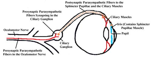
The ciliary ganglion story - after the synapse
The presynaptic parasympathetic fibers in the motor root synapse, and then the postsynaptic parasympathetic fibers unite with the fibers in the other two roots, sensory and sympathetic, to form short ciliary nerves. These short ciliary nerves travel anteriorly to reach the back of the eyeball, pierce it, and pass forward within the walls of the eyeball to reach the smooth muscle of the eyeball. The parasympathetic fibers innervate 2 muscles: the ciliary muscle, which relaxes the suspensory ligament of the lens and allows the lens to thicken for close-up vision, and the sphincter pupillae, circularly oriented fibers in the iris that constrict the pupil to allow less light to enter the eye. The sympathetic fibers innervate one muscle: dilator pupillae, radially oriented fibers in the iris that open the pupil to allow more light into the eye.
There is one more muscle in the orbit that receives sympathetic innervation: the superior tarsal muscle, which is smooth muscle near the anterior attachment of the levator palpebrae superioris muscle to the superior tarsal plate. This muscle is innervated by sympathetic fibers from the internal carotid plexus, and it holds the eyelid up. Horner's syndrome, loss of sympathetic innervation in the head, will cause ptosis, or a drooping eyelid, along with a constricted pupil and a flushed, dry face on the affected side.
|