|
|
|
||||||||||||
Dissector Answers - Hip & Posterior Thigh |
|||||||||||||
Learning Objectives:
Upon completion of this session, the student will be able to:
- Describe the anatomy of the lateral femoral (hip) region, including the gluteal muscles, their nerve supply, and their actions in locomotion.
- Identify the sacral plexus, its general plan, and its major branches in the hip and posterior thigh regions.
- Describe the muscular anatomy of the posterior thigh, its muscles, their nerve supply, and their actions in locomotion.
- Predict the functional loss and cutaneous areas affected by a given nerve injury to the hip and posterior thigh region; or conversely, given a functional and/or cutaneous loss, be able to predict which nerve or nerves are involved and the probable level of the injury.
- Define the popliteal fossa and give the spatial relationships of its contents.
- Recall the general plan of the collateral circulation at the hip and knee.
Learning Objectives and Explanations:
1. Describe the anatomy of the lateral femoral (hip) region, including the gluteal muscles, their nerve supply, and their actions in locomotion. (W&B 588-590, N353, N398, N402, N411, N490, N491, N493, N495A, N495B, N497, N499, N502, N503, N512, TG3-26A, TG3-26B, TG3-27A, TG3-27B, TG3-28)2. Identify the sacral plexus, its general plan, and its major branches in the hip and posterior thigh regions. (W&B 559-561, N499A, N499B, TG3-25A, TG3-25B, TG3-28, TG3-29)
Muscle Origin Insertion Action Innervation gluteus maximus posterior gluteal line, posterior sacrum and coccyx, sacrotuberous ligament upper fibers: iliotibial tract; lowermost fibers: gluteal tuberosity of femur extends thigh and trunk, laterally rotates thigh inferior gluteal nerve gluteus medius ilium between posterior and anterior gluteal lines greater trochanter of femur abducts and medially rotates thigh superior gluteal nerve gluteus minimus ilium between anterior and inferior gluteal lines greater trochanter of femur abducts and medially rotates thigh superior gluteal nerve tensor fasciae latae anterior part of iliac crest, anterior superior iliac spine iliotibial tract flexes, abducts, and medially rotates the thigh superior gluteal nerve piriformis anterior surface of sacrum upper border of greater trochanter of femur laterally rotates and abducts thigh ventral rami of S1-S2
obturator internus margin of obturator foramen and internal surface of obturator membrane greater trochanter, medial surface above the trochanteric fossa laterally rotates and abducts thigh nerve to obturator internus (L5,S1-2) gemellus, superior ischial spine obturator internus tendon laterally rotates the thigh nerve to obturator internus gemellus, inferior ischial tuberosity obturator internus tendon laterally rotates the thigh nerve to quadratus femoris quadratus femoris lateral border of ischial tuberosity quadrate line below intertrochanter crest laterally rotates thigh nerve to quadratus femoris 3. Describe the muscular anatomy of the posterior thigh, its muscles, their nerve supply, and their actions in locomotion. (W&B 595-597, N492, N495, N502, N540, TG3-29)Images from "Anatomy of the Human Body" by Henry Gray are provided by:
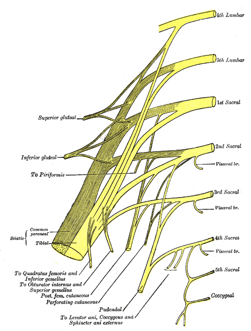
The sacral plexus proper includes contributions from L4 through part of S4. The contributions from L4 and L5 fuses to form the lumbosacral trunk, because it crosses the pelvic brim to join the sacral ventral primary rami. (Also shown here is the coccygeal plexus, which includes the rest of S4, S5, and the coccygeal nerve.) All of the contributing nerves, except S4, divide into anterior (shown yellow above) and posterior (shown yellow with black shading above) branches, but we will not dwell on the anterior/posterior division scheme. Focus instead on spinal cord segments contributing to various nerves.
sciatic nerve: actually two separate nerves that are usually fused together (though in some cases they are not) - the tibial nerve, from anterior branches of L4 through S3, and the common fibular (peroneal) nerve, from the posterior branches of L4 through S2. The sciatic nerve runs under the piriformis muscle, emerging at its inferior border. It travels through the greater sciatic foramen, and on into the thigh. The sciatic nerve provides motor innervation to the flexors of the knee (posterior thigh muscles) and all of the muscles in the leg and the foot.
superior gluteal nerve: from posterior branches of L4 through S1. Travels through the greater sciatic foramen superior to the piriformis muscle, and moves laterally between the gluteus medius muscle and the gluteus minimus muscle. Provides motor innervation to gluteus medius muscle, gluteus minimus muscle, and tensor fascia lata muscle.
inferior gluteal nerve: from the posterior branches of L5 through S2. Travels through the greater sciatic foramen inferior to the piriformis muscle, and travels for a short distance deep to the gluteus maximus muscle but superficial to the sciatic nerve. It provides the sole motor innervation of the gluteus maximus muscle.
nerve to quadratus femoris muscle: from the anterior branches of L4 through S1. Travels through the greater sciatic foramen inferior to the piriformis muscle. Provides motor innervation to quadratus femoris and inferior gemellus muscles.
nerve to obturator internus muscle: from the anterior branches of L5 through S2. Travels through the greater sciatic foramen inferior to the piriformis muscle, but then loops back around to reenter the pelvis through the lesser sciatic foramen. Provides motor innervation to the obturator internus and superior gemellus muscles.
posterior femoral cutaneous nerve: from the anterior branches of S2 and S3 and the posterior branches of S1 and S2. Travels through the greater sciatic foramen inferior to the piriformis muscle, and lies alongside the sciatic nerve. It provides sensory innervation to the skin of the back of the thigh in addition to the skin of the lower and lateral buttocks (through gluteal branches) and the perineum (via perineal branches).
pudendal nerve: from the anterior branches of S2 through S4. Travels through the greater sciatic foramen inferior to the piriformis muscle, but then loops back around to enter the perineum through the lesser sciatic foramen, entering the pudendal canal. Provides motor innervation to the muscles of the perineum, and is the primary sensory innervation to the genitalia. (Latin, pudere = to be ashamed)
perforating cutaneous nerve: from the posterior branches of S2 and S3. Pierces the sacrotuberal ligament to provide sensory innervation to the skin of the medial part of the fold of the buttock.
nerve to piriformis muscle: from posterior branches of S1 and S2. Provides motor innervation to the piriformis muscle.
nerves to coccygeus and levator ani muscles: from anterior branches of S3 and S4. Provide motor innervation to coccygeus and levator ani muscles.
pelvic splanchnic nerves: from the anterior branches of S2 through S4. Travel to the inferior hypogastric plexus (see below).
perineal branch of the fourth sacral nerve: from the anterior branch of S4. Descends through the coccygeus muscle and runs anteriorly to the external sphincter ani muscle. Provides motor innervation to this muscle and sensory innervation to the overlying skin.
4. Predict the functional loss and cutaneous areas affected by a given nerve injury to the hip or posterior thigh region; or conversely, given a functional and/or cutaneous loss, be able to predict which nerve or nerves are involved and the probable level of the injury.The posterior thigh contains the hamstring muscles - the semitendinosus, semimembranosus, and biceps femoris muscles. They all arise on the ischial tuberosity and cross the knee to insert. The short head of the biceps femoris muscle is not a hamstring muscle, since it arises from the back of the femur, its innervation is different than the rest. See the table below.
Muscle Origin Insertion Action Innervation biceps femoris long head: ischial tuberosity; short head: lateral lip of linea aspera head of fibula and lateral condyle of the tibia extends thigh, flexes leg long head: tibial nerve; short head: common fibular (peroneal) nerve semitendinosus lower, medial surface of ischial tuberosity (common tendon with biceps femoris m.) medial surface of tibia (via pes anserinus) extends thigh, flexes leg tibial nerve semimembranosus upper, outer surface of ischial tuberosity medial condyle of tibia extends thigh, flexes leg tibial nerve Note: There is also a hamstring portion of the adductor magnus muscle which inserts on the adductor tubercle and is innervated by the tibial nerve.5. Define the popliteal fossa and give the spatial relationships of its contents. (N493, N495, N502, N512, N517, N546, TG3-31A, TG3-31B)
- Example #1: If the superior gluteal nerve (L4, L5, and S1) is injured, the muscles affected are the gluteus medius, gluteus minimus, and tensor fasciae latae. These muscles stabilize the pelvis when we walk, preventing our hips from dropping as we lift a foot off the ground to propel forward. The pelvis drops on the opposite side of the nerve injury. In other words, the right gluteus medius and gluteus minimus support the pelvis in such a way that when the left foot is lifted off the ground, the left pelvis remains level with the right pelvis, but when the right gluteal muscles are injured, the left side of the pelvis will drop when the left foot is lifted off the ground.
- Example #2: damage to the femoral nerve (L2, L3, and L4) impairs actions of sartorius, iliopsoas, and quadriceps muscles. This will result in weak or absent flexion of the thigh and extension of the knee.
- Example #3: damage to the inferior gluteal nerve (L5, S1, and S2) impairs the gluteus maximus muscle. This will result in weak or absent extension of the thigh.
- Example #4: damage to the sciatic nerve (L4 through S3) will impair the action of the hamstrings. This will result in weak or absent extension of the thigh and/or flexion of the knee. Furthermore, the leg and foot will lose innervation, resulting in foot drop.
- Example #5: damage to the obturator nerve (L2, L3, and L4) will impair the action of the adductors and obturator externus muscle. This will result in weak or absent adduction and medial rotation of the thigh. (OTE: The obturator externus muscle is a lateral rotator, but there are other muscles that do that, so it would not be missed).
6. Recall the general plan of the collateral circulation at the hip and knee. (W&B 652-654, TG3-27B, TG3-38, TG3-62)The popliteal fossa is defined superomedially by the tendons of the semimembranosus and semitendinosus muscles, superolaterally by the tendon of the biceps femoris muscle, and inferiorly by the medial and lateral heads of the gastrocnemius muscle. Within the fossa lie the popliteal artery and vein, including branches and tributaries, and the tibial and common fibular nerves. (Greek, gastrokneme = calf of the leg)
In the popliteal fossa, the popliteal artery lies against the back of the knee joint. The popliteal vein is posterior to the artery (more superficial). The common fibular nerve descends toward the fibular neck, while the tibial nerve bisects the fossa and lies posterior to popliteal vein. So, we have nerve, vein, and artery going posterior (superficial) to anterior (deep).The "cruciate anastomosis" of the thigh involves links between the inferior gluteal artery (from the internal iliac artery) and the medial circumflex femoral artery , lateral circumflex femoral artery, and first perforating branch of the deep femoral artery. (These all arise from the deep femoral artery, which is a branch of the femoral artery, which is the continuation of the external iliac artery). There are also other anastomoses between branches of the external iliac artery and branches of the internal iliac artery, often via the arteries of the abdominal wall. All of these can help supply blood to the lower limb in the event of an obstruction of the external iliac artery.
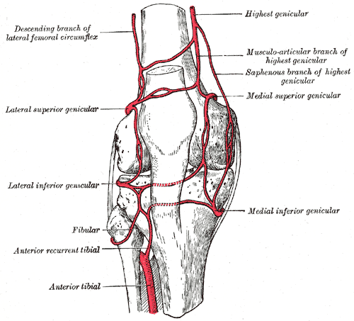
The "genicular anastomosis" in the region of the knee is also very important. It involves ten vessels. Two from above (the descending branch of the lateral circumflex femoral artery and the descending genicular branch (highest genicular) of the femoral artery), five from the popliteal artery, and three from below, usually branches of the anterior tibial artery. This system can help supply blood to the leg in the event of an obstruction of the femoral artery below the branching-off of the deep femoral artery.
Cultural enrichment: Check out these sections from the 1918 version of Gray's Anatomy of the Human Body! Some of the terms are (of course) out-of-date, but the illustrations are timeless. The Femur - The Muscles and Fasciae of the Thigh - The Veins of the Lower Extremity, Abdomen, and Pelvis - The Arteries of the Lower Extremity - Surface Anatomy of the Lower Extremity - Surface Markings of the Lower Extremity
Questions and Answers:
1. What nerves accompany the lesser saphenous vein?The medial sural cutaneous nerve and sural nerve accompany the lesser saphenous vein. N538,TG3-032. Where does the lesser saphenous vein disappear? Where does it terminate?The lesser saphenous vein pierces the popliteal fascia covering the popliteal fossa at the back of the knee. In that area it drains into the popliteal vein. N545,TG3-03,TG3-333. How is the sural nerve formed?The sural nerve is formed from fibers contributed by both the tibial and common fibular nerves. N545,TG3-034. Where does the sural nerve distribute and by what name?It distributes to the posterolateral aspect of the lower leg, the ankle, and the heel. Its continuation on the lateral side of the foot, all the way down to the little toe, is called the lateral dorsal cutaneous nerve. N545,TG3-03,TG3-445. Note that both gluteus maximus and tensor fasciae latae insert via the iliotibial tract of the fascia lata. Where does the iliotibial tract attach? (TG3-57)6. Locate the superior gluteal artery and nerve coursing deep to the gluteus medius muscle. What are their sources?Superiorly, the iliotibial tract attaches to the iliac tubercle. Inferiorly, it attaches to the lateral condyle of the tibia.
7. What is the innervation and blood supply to the gluteus maximus muscle?The superior gluteal artery originates from the posterior division of the internal iliac artery. The superior gluteal nerve is from the sacral plexus (L4-S1). It passes through the greater sciatic foramen above the piriformis muscle, then passes between the gluteus medius and minimus muscles to innervate them and the tensor fascia latae muscle. (TG3-25, TG6-17)
8. Locate the piriformis muscle and trace it to its insertion. Through what foramen does it exit the pelvis?The inferior gluteal nerve innervates gluteus maximus, while the superior and inferior gluteal arteries supply it with blood. (TG3-26)
9. Through what foramen does obturator internus muscle pass?The piriformis muscle exits the pelvis via the greater sciatic foramen. (TG6-22A, TG6-22C)
10. What major action do the gemelli muscles, obturator internus muscle, and the quadratus femoris muscles have in common?The obturator internus muscle exits the pelvis via the lesser sciatic foramen. (TG6-23)
11. Is there a perforating cutaneous nerve and artery associated with the sacrotuberal ligament?They are all lateral rotators of the femur.
12. What structures do the greater and lesser sciatic foramina transmit?A perforating cutaneous neurovascular bundle usually passes through the sacrotuberal ligament to reach the inferomedial portion of the buttock.
13. Do you find a branch from the tibial division of the sciatic nerve to adductor magnus; and one from the common fibular division to the short head of biceps?Greater Sciatic Foramen transmits:
- piriformis muscle
- superior and inferior gluteal vessels and nerves
- internal pudendal vessels and pudendal nerve**
- sciatic nerve
- posterior femoral cutaneous nerve
- nerves to the obturator internus** and quadratus femoris mm.
Lesser Sciatic Foramen transmits:
- tendon of the obturator internus m.
- nerve to the obturator internus**
- internal pudendal vessels and pudendal nerve**
**pass through both the greater and lesser sciatic foramen (TG3-26)
14. The popliteal fascia is part of what fascia?You should! (TG3-29AB)
15. Where exactly is the popliteal fossa located in the lower extremity?The popliteal fascia is continuous with fascia lata above, and crural fascia below. (TG3-03)
16. Do you find lymph nodes in the popliteal fossa?See #7 above. (TG3-31)
A few popliteal nodes lie embedded in the fat there. They receive lymph following the lesser saphenous vein. (TG3-70)