|
|
|
||||||||||||
Dissector Answers - Female Reproductive Anatomy |
|||||||||||||
Learning Objectives:
Upon completion of this session, the student will be able to:
- Trace the skeletal and ligamentous boundaries of the perineum, and define the anal and urogenital triangles.
- Identify the superficial features of the external genitalia in the female.
- Describe the structure, contents, and course of the pudendal canal.
- Trace the branching pattern of the internal pudendal vessels and the pudendal nerve.
- Differentiate between male and female urethrae.
- Identify the components of the external genital organs and give the homologues in each of both sexes.
- Describe structure and function of the erectile bodies.
- Identify the muscles and fasciae of the perineum and their functions.
- Trace the nerve and blood supply to the external genital organs and the muscles of the perineum.
- Trace the lymphatic drainage of the perineum.
- Trace the continuity of the abdominal peritoneum with that of the pelvic cavity, and identify the peritoneal pouches of the pelvic floor in both sexes.
- Describe the relationships of the bladder to other pelvic organs in both sexes.
- Describe the normal position and relationships of the organs of the female reproductive tract and the role of each in reproductive processes.
- Describe the broad ligament and differentiate its parts.
- Identify the ovary and discuss the functional significance of its ligaments.
- Demonstrate the uterine tube and its subdivisions.
- Identify the uterus and its subdivisions and demonstrate the continuity of its lumen with that of the uterine tubes and the vagina.
- Differentiate between the internal and external os of the cervix.
- Identify the vagina, and note the angle formed at its junction with the uterus.
- Describe the support mechanisms for the uterus which act to prevent uterine prolapse.
- Describe the formation of the two sciatic foramina. List the muscles, nerves, and vessels which pass through each.
- Describe the general gross features of the breast and its blood supply, innervation, and lymphatic drainage.
Learning Objectives and Explanations:
1. Trace the skeletal and ligamentous boundaries of the perineum, and define the anal and urogenital triangles. (W&B 519, N 379A, 379B, TG 6-24A, 6-24B)2. Identify the superficial features of the external genitalia. (W&B 519-522, N 351, 377, 382, 387, 390, 398, TG 6-02, 6-25A, 6-25B, 6-31)Officially, the perineum is the outlet of the pelvis. (Used more loosely, it can refer to the area of skin between the anus and the posterior part of the external genitalia.) It is diamond-shaped, and can therefore be divided into two isosceles triangles by a line drawn between the ischial tuberosities. The anterior, or urogenital triangle has as its apex the pubic symphysis, with the ischiopubic rami as equal sides, and our imaginary line as the base. The posterior, or anal triangle is upside-down, with our line again as the base, the sacrotuberal ligaments as the equal sides, and the coccyx as the apex. (peri + inan ("to empty out" in Greek))
You really need to think about this in 3-D because, although drawn two-dimensionally from an inferior point of view it looks like the coccyx, anus, vagina, and pubic symphysis are all coplanar, they are not.
Sorry to cop out and paste in the tables, but there is little connectivity or functionality that needs to be explained. It is pretty much a "these are the things you should know about" situation.
3. Describe the structure, contents, and course of the pudendal canal. (W&B 524-526, N 404, 405, 411, 413, TG 6-28A, 6-28B)4. Trace the branching pattern of the internal pudendal vessels and the pudendal nerve. (W&B 524-526, N 404, 405, 411, 413, TG 6-29A, 6-29B, 6-30A, 6-30B)The pudendal canal extends from the lesser sciatic foramen, where its contents enter the perineum, to the posterior edge of the perineal membrane. It contains the internal pudendal artery, internal pudendal vein, and the pudendal nerve.
5. Differentiate between male and female urethrae. (W&B 542-543, N 369, 379, 384, 385, TG 6-08A, 6-08B, 6-09A, 6-09B, 6-10A, 6-10B)The internal pudendal artery gives off the following branches. The internal pudendal vein receives analogous tributaries.
- Within pudendal canal:
- inferior rectal artery: supply to lower rectum and anus
- perineal artery: supply to bulbospongiosus muscle and ischiocavernosus muscles, as well as posterior scrotal or posterior labial artery to supply the skin of the respective structures
- Within urogenital triangle:
- artery of the bulb of the vestibule (in females) or artery of the bulb of the penis (in males): supply to respective structures
- deep (central) artery of clitoris (in females) or deep (central) artery of the penis (in males) - within corpus cavernosum of appropriate structure
- dorsal artery of clitoris (in females) or dorsal artery of penis (in males): runs entire length of appropriate structure, sending branches to corpus cavernosa and terminate in branches to glans and prepuce.
The pudendal nerve has the following branches:
- Within pudendal canal:
- inferior rectal nerves: supply to external sphincter ani muscle and skin of anus
- perineal nerve: gives off posterior labial nerve (in female) or posterior scrotal nerve (in male), which supplies the skin of the perineum - also gives off the deep perineal nerve, which supplies motor innervation to all of the muscles of the urogenital triangle.
- Within urogenital triangle:
- dorsal nerve of clitoris (in female) or dorsal nerve of penis (in male): supply to appropriate structure
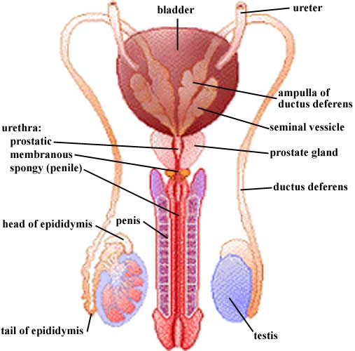
6. Identify the components of the external genital organs and give the homologues in each of both sexes. (W&B 543-551, 403-413, N 379A, 379B, 382A, 382B, 418A, 418B, TG 6-25A, 6-25B, 6-27A, 6-27B)The female urethra is about four centimeters long and is entirely "membranous urethra". The male urethra is longer than the female urethra, and is divided into three parts: membranous, prostatic, and penile (spongy) urethra. (See Pelvis & Pelvic Viscera Dissector Answers.)
7. Describe structure and function of the erectile bodies. (W&B 520-523, 527-528, N 379A, 379B, 381, 382A, 382B, TG 6-27A, 6-27B, 6-32)
Female Male vestibular bulbs corpus spongiosum greater vestibular glands bulbourethral glands urethral and paraurethral glands prostate gland glans clitoris glans penis prepuce of clitoris prepuce of penis corpus of clitoris corpus (shaft) of penis labia minora penoscrotal raphe labia majora scrotum 8. Identify the muscles and fasciae of the perineum and their functions. (W&B 524-525, N 404, 405, 411, 413, TG 6-29, 6-30A, 6-30B)The three primary erectile bodies of the penis are the two corpora cavernosa and the corpus spongiosum. These structures are surrounded by a dense tunica albuginia such that, when they are engorged with blood, the penis becomes erect. The glans penis, the expanded cap of the corpus spongiosum, remains more malleable during erection because it has a much thinner tunica albuginea than the rest of the components of the penis.
The corpora cavernosa are rooted in the perineum by two crura of the penis. Each crus is attached to the ischiopubic ramus. The corpus spongiosum is rooted as the bulb of the penis, which is attached to the perineal membrane, and receives the membranous urethra as it transverses the membrane.
There are homologous structures in the female. The clitoris contains corpora cavernosa as its erectile tissue. Like in the male, each crus is attached to the ischiopubic ramus and perineal membrane. Instead of a corpus spongiosum, the female has two vestibular bulbs, which lie along the sides of the vestibule, and also expand as the glans clitoris to cap the distal ends of the corpora cavernosa.
9. Trace the nerve and blood supply to the external genital organs and the muscles of the perineum. (W&B 524-525, N 404, 405, 411, 413, TG 6-29, 6-30A, 6-30B)The perineal membrane stretches between the two ischiopubic rami and serves as an anchor for the erectile bodies and their associated muscles. It also divides the perineum into superficial and deep spaces, sometimes referred to as "pouches". The perineal membrane is set at an angle (as are the ischiopubic rami), so that the superficial perineal pouch is anteroinferior to the perineal membrane while the deep perineal pouch is posterosuperior to the perineal membrane. The best way to consider these structures is to look at the tables:
perineal membrane (N379, N381, TG6-27A, TG6-27B) membrane stretching across the urogenital triangle attaching to both ischiopubic rami perineal membrane is pierced by the urethra, vagina and branches of the pudendal neurovascular bundle perineal body (N379, TG6-27A) fibrous connective tissue in the midline between the vestibule of the vagina and the anus many of the muscles of the perineum attach at or to the perineal body, including the transverse perineal muscles, bulbospongiosus, and external anal sphincter; the male version is often called the central tendinous point
Muscles
Muscle Origin Insertion Action Innervation Artery Notes Image bulbospongiosus, in female (N379, N382, TG6-27A, TG6-27B) perineal body and fascia of the bulb of the vestibule perineal membrane and corpus cavernosum of the clitoris compresses the vestibular bulb and constricts the vaginal orifice deep branch of the perineal nerve (from pudendal nerve) perineal a. skeletal muscle ischiocavernosus (N379, N382, TG6-27A, TG6-27B) medial surface of the ischial tuberosity and the ischiopubic ramus corpus cavernosum and crus of the penis/clitoris compresses the corpus cavernosum deep branch of the perineal nerve (from pudendal nerve) perineal a. ischiocavernosus m. is closely applied to the surface of the crus penis/clitoris sphincter urethrae, in female (N379, N382, N385, TG6-28A, TG6-28B) encircles the urethra encircles urethra and vagina; extends superiorly along the urethra as far as the inferior surface of the bladder compresses urethra and vagina deep branch of perineal nerve from pudendal nerve internal pudendal a. skeletal muscle (Greek, sphincter = that which binds tight) superficial transverse perineus medial surface of the ischial ramus contralateral muscle and the perineal body/central tendinous point fixes and stabilizes perineal body/central tendinous point deep branch of perineal nerve from pudendal nerve perineal a. superficial and deep transverse perineus muscles are separated by the perineal membrane deep transverse perineus medial surface of the ischial ramus contralateral muscle and perineal body/central tendinous point fixes and stabilizes the perineal body/central tendinous point deep branch of perineal nerve from pudendal nerve internal pudendal a. superficial and deep transverse perineus muscles are separated by the perineal membrane
10.Trace the lymphatic drainage of the perineum. (N 406, 407, 408A, 408B, TG 6-33, 6-34)The nerve supply to the external genital organs is via the pudendal nerve, which gives off the dorsal clitoral or penile nerve, which is sensory for the clitoris/penis, and also gives off the perineal nerve, which in turn gives off posterior labial or posterior scrotal nerves (sensory) and the deep perineal nerve supplying all of the muscles of the urogenital triangle.
The autonomic supply to the erectile bodies arrives via the cavernous nerves from the inferior hypogastric plexus, which descend lateral to the prostate (male) or membranous urethra (female) to reach the erectile tissue of the corpus cavernosum and corpus spongiosum. The parasympathetic fibers are responsible for relaxing the vascular smooth muscle of the deep arteries and arteries of the bulb, allowing blood to fill the erectile tissue, resulting in erection.
The internal pudendal artery gives off the dorsal artery of the clitoris or penis and the deep artery of the clitoris or penis as terminal branches. It also gives off an artery to the bulb of the vestibule (in females) or an artery to the bulb of the penis (in males). The perineal artery gives off a posterior labial or posterior scrotal artery.
11. Trace the continuity of the abdominal peritoneum with that of the pelvic cavity, and identify the peritoneal pouches of the pelvic floor in both sexes. (W&B 533-534, N 360, 361, 362, 363, 371, TG 6-07A, 6-07B, 6-08A, 6-08B, 6-11, 6-13)
- perineum and external genitalia: drain to superficial inguinal nodes
- anal canal: superior part drains to internal iliac nodes, inferior part drains to superficial inguinal nodes
- testes: lymphatic vessels run with the spermatic cord, terminating in the lumbar nodes
12. Describe the relationships of the bladder to other pelvic organs in both sexes. (W&B 533-534, N 360, 361, 362, 366, 402, 403, 406, 407, 408, 410, TG 6-07A, 6-07B, 6-08A, 6-08B)The peritoneum continues from the abdominal cavity into the pelvic cavity, but does not entirely invest the pelvic viscera. In the female, the peritoneum:
- extends from the anterior abdominal wall to the superior surface of the bladder, not drooping low enough to catch the anterior surface
- sweeps over the fundus and covers part of the posterior surface of the bladder
- jumps from the posterior surface of the bladder to the anterior (vesicle) surface of the uterus
- sweeps superiorly, to the fundus of the uterus, contacting the uterine tubes
- the space created by the peritoneum sweeping down the back of the bladder, over to the uterus, and up the front of the uterus is the vesicouterine pouch
- continues around the fundus of the uterus, and over the uterine tubes, to the posterosuperior (intestinal) surface of the uterus
- the "doubling" of the peritoneal layers as they hang on either side of the uterine tubes creates the broad ligaments
- jumps from the uterus to cover the anterior portion of the rectum, starting about 2/3 of the way down the rectum
- continues up the rectum, investing the sides as well as it reaches the superior 1/3, attaching to the posterior body wall
- the space created by the peritoneum sweeping across the uterus, jumping to the rectum, and beginning to travel up the front of the rectum is the rectouterine pouch
13. Describe the normal position and relationships of the organs of the female reproductive tracts and the role of each in reproductive processes. (W&B 543-553, N 360, 362, 369, 370, 371A, 371B, 378, 382A, 382B, 383, 399, 400, 402, 404A, 404B, 352, 359, 361A, 361B, 362, 363, 365, 384A, 384B, TG 5-34, 6-07A, 6-07B, 6-08A, 6-08B, 6-09A, 6-09B, 6-10A, 6-10B, 6-11, 6-12, 6-14, 6-15, 6-17, 6-23, 6-29, 6-31)The bladder lies in the anterior half of the pelvis, bounded anteriorly and laterally by the pubic symphysis. Posterior to it we have:
- female: vesicouterine septum (pouch), vagina, and uterus (also somewhat superior to the bladder)
- male: rectovesicular septum (pouch), rectum, ductus deferens, and seminal vesicles
Inferior to the bladder we find the pelvic diaphragm (in females) or the prostate gland (in males).
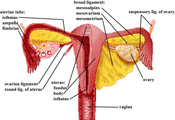
14. Describe the broad ligament and differentiate its parts. (W&B 547-548, N 371, TG 6-07, 6-08, 6-11, 6-12)These diagrams will be useful here, and for the remaining objectives in this session:
Female:
Male:
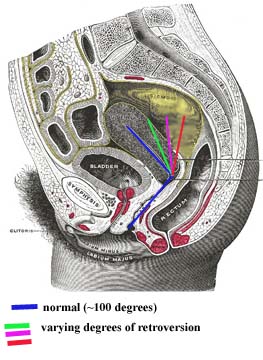
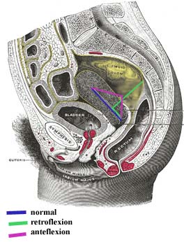
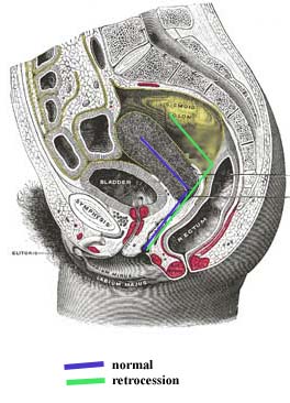
Female: The uterus is more or less horizontal in orientation. It actually lies upon the bladder, so its vesical surface is partially anterior but mostly inferior. Its intestinal surface faces the pelvic cavity. Deviations from this "flopped over the bladder" position can happen. Terms that are "________version" refer to the angle between the long axis of the uterus and the long axis of the vagina. This angle is usually about 100 degrees. Cases where the uterus stands more vertically, and therefore that angle approaches 180 degrees, are called RETROversion. Terms that are "________flexion" refer to the long axis of the body of the uterus as compared to the long axis of the uterine cervix. The long axis of a normal body of the uterus is relatively horizontal, while in the cervix the axis turns partially vertical. With RETROflexion the uterus is actually flopped back, away from the bladder. With ANTEflexion, the uterus is kinked, with a marked angle between the long axis of its body and the long axis of its cervix. (A slight bit of anteflexion is very normal.) Cases where the entire uterus, cervix and all, is moved posteriorly are called retrocession. The vagina is longer here.
Images from "Anatomy of the Human Body" by Henry Gray are provided by:
The uterine (fallopian) tubes attach somewhat laterally to the uterus. They reach toward the ovaries, but are not "officially" attached to them. The fimbriae of the uterine (fallopian) tubes come closest to contacting the ovary. The ovaries themselves are the female gonads. They lie against the pelvic walls.
The female gametes (ova, eggs) mature in the ovaries. In the normal case, one egg is released per month from one ovary or the other. This egg is actually released into the pelvic cavity, but the waving motion of the fimbriae help guide the egg into the uterine tube. The tube is the usual site of fertilization of the egg by sperm, then the newly formed zygote travels to the uterus. It is the job of the uterus to provide an environment for growth of the embryo.
15. Identify the ovary and discuss the functional significance of its ligaments. (W&B 547-548, N 369, TG 6-07, 6-08, 6-11, 6-12)The broad ligament is a section of peritoneum, like a mesentery, which extends from the pelvic walls to the uterus and uterine (fallopian) tubes. Its three parts are continuous with each other, making it difficult to discern at their junctions. The mesosalpinx is the peritoneum that covers the uterine tube and hangs below it to meet with the mesovarium. The mesovarium is the peritoneum covering the ovary and ovarian ligament, extending like a shelf posteriorly from the mesosalpinx. (If you squint at the diagram in #11, you'll see a ridge of peritoneum running along the top of each ovary.) The mesometrium is the rest of the broad ligament - all of the peritoneum directly connected to the uterus and extending toward the lateral abdominal wall. (Greek, metra = uterus, from meter (mother);
16. Demonstrate the uterine tube and its subdivisions. (W&B 547-548, N 371, TG 6-07, 6-08, 6-11, 6-12)The suspensory ligaments of the ovaries are peritoneal folds covering the ovarian neurovascular supply as the vessels pass over the pelvic brim and into the pelvis to reach the ovary. The suspensory ligament conducts ovarian arteries and veins, nerves, and lymphatics to the ovary. The "ovarian ligament" proper is a round cord which attaches the ovary to the uterus just below the entrance of the uterine tube into the uterus. The ovarian ligament, a remnant of a portion of the gubernaculum, is within the mesovarium.
17. Identify the uterus and its subdivisions and demonstrate the continuity of its lumen with that of the uterine tubes and the vagina. (W&B 548-551, N 371, TG 6-07, 6-08, 6-11, 6-12)The uterine tube extends laterally about 10 cm from the uterus to the ovary. It has three parts:
- isthmus: the constricted part adjacent to the uterus
- ampulla: the widest and longest part, extending laterally to the infundibulum from the isthmus
- infundibulum: the funnel-like terminus, with fringed processes called fimbriae that contact the ovary.
18. Differentiate between the internal and external os of the cervix. (W&B 551-553, N 371, TG 6-11, 6-12)The lumen of the uterus is continuous on both sides with the lumens of the uterine tubes. It is continuous inferiorly with the lumen of the vagina. It is divided into four parts:
19. Identify the vagina, and note the angle formed at its junction with the uterus. (W&B 551-553, TG 6-08, 6-11, 6-12, 6-13)The tapered neck or cervix of the uterus is traversed by the cervical canal. Above, it is continuous with the cavity of the body of the uterus at the internal os. Below, at a depression on the vaginal portion of the cervix, the external os opens into the cavity of the vagina.
20. Describe the support mechanisms for the uterus which act to prevent uterine prolapse. (W&B 551-553, TG 6-08, 6-11, 6-12, 6-13)The vagina is muscular, but not as much as the uterus. The angle between its long axis and the long axis of the uterus is about 100 degrees in normal cases. Other cases can occur, as described in #12 above.
21. Describe the formation of the two sciatic foramina. List the muscles, nerves, and vessels which pass through each. (W&B 570-571, N 352, 503, TG 3-28, 6-06)There are multiple mechanisms and structures that help to support the uterus. As described above, the normal position of the uterus is anteverted superior to the bladder, and anteflexed at the isthmus. This tends to bring the uterus to rest upon the superior surface of the bladder, thereby helping to support its weight and to prevent uterine prolapse (descent of the uterus down the vagina).
Although there are various opinions about the quality of uterine support from various mechanisms, most would agree that the single most important mechanism for uterine support is the pelvic diaphragm. Strength and integrity of the pelvic diaphragm is essential for maintaining the proper positions of all pelvic viscera.
In addition to the pelvic diaphragm, there are various ligaments that are individually weak but when added together help to maintain proper uterine position. The broad ligament, which is the peritoneum covering the uterus, uterine tubes, and ovaries, helps to maintain their relative positions. Beneath the anterior leaf of the mesometrium, the round ligament of the uterus pull toward the deep inguinal ring and helps to maintain the uterus in its anteverted position. At the base of the broad ligament, condensations of endopelvic connective tissue form what are referred to as ligaments which attach primarily to the cervical region of the uterus and help to support this portion of the uterus. These ligaments are the pubocervical ligament (attaching anteriorly to pubis), transverse cervical or cardinal ligament (attaching to the lateral pelvic wall and surrounding the uterine vessels), and the uterosacral ligament (attaching posteriorly to sacrum, obviously, and containing some smooth muscle).
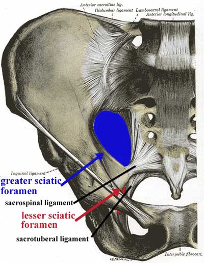
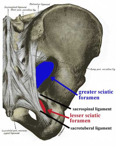
22. Describe the general gross features of the breast and its blood supply, innervation, and lymphatic drainage. (W&B 107-110, image, 182A, 182B, 184, TG 2-10A, 2-10B, 2-11A, 2-11B)This is something that only makes sense in three dimensions. You must look at a pelvis from your bone box or in the lab, and figure out where these ligaments are going. Luckily, they are named in a very logical way. These anterior and posterior views might help.
Images from "Anatomy of the Human Body" by Henry Gray are provided by:
The sacrotuberal ligament connects the sacrum to the the ischial tuberosity. With the pelvis in the correct position, it runs mostly inferolaterally from the sacrum to the tuberosity, and only slightly anteriorly. The sacrospinal (sacrospinous) ligament connects the sacrum to the ischial spine. With the pelvis in correct anatomical position, it runs anterolaterally from the sacrum to the ischial spine, but does not deviate much in the superior-inferior axis.
These ligaments, along with the greater sciatic notch and the lesser sciatic notch, make up the greater sciatic foramen and the lesser sciatic foramen, respectively.
greater sciatic foramen: bounded anteriorly and superiorly by the posterior border of the hip bone (greater sciatic notch), posteriorly by the sacrotuberal ligament, and inferiorly by the sacrospinal ligament. The piriformis muscle passes through this opening, as do these nerves and vessels:
- superior to piriformis muscle: superior gluteal vessels and nerve
- inferior to piriformis muscle: inferior gluteal vessels and nerve, the sciatic nerve, the posterior femoral cutaneous nerve, and the nerve to the quadratus femoris muscle - also, the internal pudendal vessels and nerve and the nerve to the obturator internus muscle leave the pelvis via this opening, but enter the perineum through the lesser sciatic foramen (see below)
lesser sciatic foramen: bounded anteriorly by the ischial tuberosity, superiorly by the ischial spine and sacrospinal ligament, and posteriorly by the sacrotuberal ligament. It transmits the tendon of the obturator internus muscle - also, the nerve to the obturator internus muscle and the internal pudendal vessels and nerve, which left the pelvis via the greater sciatic foramen, re-enter the pelvis (in the case of the nerve to the obturator internus muscle) or the perineum (in the case of the internal pudendal vessels and nerve) via the lesser sciatic foramen
The mammary gland is a modified sweat gland. It is entirely contained within subcutaneous tissue. The most important internal gross features are glandular, namely secretory glands, lactiferous ducts, and lactiferous sinuses. (It is the glandular nature of the breast that makes it a common site for the development of cancer.) Externally, an important feature is the nipple, which is surrounded by the areola. Each of the approximately 20 lactiferous sinuses have an individual opening on the nipple.
The breast's arterial supply is derived from branches of the internal thoracic artery (including anterior intercostals), the lateral thoracic artery, the thoracoacromial artery, and posterior intercostal arteries. Venous drainage follows arterial supply, primarily draining into the axillary vein, but also draining some blood into the internal thoracic vein.
Lymph passes from the nipple, areola, and lobules to the subareolar lymphatic plexus. From there:Lymphatic vessels in the skin of the breast drain into the axillary, inferior deep cervical, infraclavicular, and parasternal lymph nodes.
- MOST (75%) of the lymph goes to the axillary lymph nodes, via the pectoral lymph nodes. (It is extremely important to consider the axillary nodes when performing a breast exam on a patient.)
- Most of the rest goes to the parasternal lymph nodes.
- A small amount of lymph goes to the opposite breast.
- A small amount of lymph goes to the abdominal wall and downward.
Lymph from the axillary lymph nodes subsequently drains into the subclavian lymph trunk. Lymph from parasternal nodes enters the bronchomediastinal trunk.
Cultural enrichment: Check out these sections from the 1918 version of Gray's Anatomy of the Human Body! Some of the terms are (of course) out-of-date, but the illustrations are timeless.
Questions and Answers:
1a. Note the difference between male and female in the subpubic angle, the angle formed by the subpubic arch. What are other sex differences in the pelvic skeleton? (W&B 571-573, N 354, TG 6-05A, 6-05B, 6-05CD, 6-05EF)2a. Do you find muscular (deep) branches of the perineal nerves? (N 411, 413, TG 6-30A, 6-30B)
Structure/Section Female Male pelvic inlet oval and rounded heart-shaped pelvic outlet large small pubic arch and subpubic angle wide narrow iliac wings flared less flared The deep perineal nerve innervates all of the muscles of the urogenital triangle via slender branches that may be difficult to locate.3a. What is the source of the deep (central) artery of the clitoris/penis? (N 404, 405, 6-29A, 6-29B)This artery is a branch of the internal pudendal artery.4a. What is the source and drainage of the deep dorsal vein of the clitoris/penis and the dorsal veins and arteries of the clitoris/penis? (N 265, 383, 359, 404, 405, 381, TG 5-34, 6-10, 6-23, 6-27, 6-29)5a. What is the function of the perineal membrane? (N 404, 405, 411, 413, TG 6-27A, 6-27B, 6-28A, 6-28B)The deep dorsal veins drain into the vesical venous plexus. The "normal" dorsal veins drain into the superficial external pudendal vein. The dorsal arteries come from the internal pudendal arteries.
This membrane covers the anterior part of pelvic outlet. It aids in support of the pelvic viscera and as an attachment for perineal structures. It is pierced by the arteries of the erectile bodies and the dorsal arteries and nerves of the clitoris or penis.6a. On the sagittally-sectioned female specimen, trace the peritoneum from the ventral abdominal wall; examining the vesicouterine pouch and its manner of reflection from the bladder to the uterus. Onto what part of uterus does it reflect? (N 378, TG 6-08A)7a. Trace the peritoneum across the uterus and define the rectouterine pouch. Note peritoneum on the posterior wall of the vagina. From what point does the peritoneum reflect to the rectum? What is the significance of this? (N 360, TG 6-08A)The peritoneum on the superior surface of the bladder reflects onto the uterus at the isthmus, just superior to the cervix.
8a. Within the broad ligament, locate the ovarian ligament and the round ligament of the uterus. Consider development and continuities of these structures. (N 367, 420, TG 6-11, 6-12)The peritoneum of the rectouterine pouch lies in contact with the posterior fornix of the vagina. This allows incisions, punctures, or lacerations of the posterior fornix of the vagina to open the peritoneal cavity. (This is often how eggs are harvested these days.)
9a. Locate and define the peritoneal fold called the suspensory ligament of the ovary. What does it contain? (N 374, 400, TG 6-11A, 6-11B, 6-12)The proper ovarian ligament and the round ligament of the uterus are both remnants of the gubernaculum. They are continuous with one another where they contact the lateral surface of the uterus inferior to the uterine tube.
10a. Strip the peritoneum from the suspensory ligament of the ovary on one side and trace the ovarian artery and vein. What are their sources? (N 400, TG 5-34)The suspensory ligament of the ovary contains ovarian vessels, autonomic nerves, lymphatics, and extraperitoneal connective tissue.
11a. What is the complete area of distribution of the ovarian artery? (N 400, TG 6-11B, 6-12)The ovarian artery branches from aorta. The right ovarian vein drains to inferior vena cava. The left ovarian vein drains to left renal vein. (This is analogous to testicular vessels in males.)
12a. Locate the ureter. Note its relation to uterine artery. Trace it to the bladder and posteriorly to the brim of the pelvis, noting course, relation to peritoneum, and blood supply. (N 400, TG 6-11B, 6-17)The ovarian artery supplies the ovary, mesovarium, and infundibulum of the uterine tube.
13a. Trace the round ligament from the uterus to the deep inguinal ring. Where does it attach? (N 369, TG 6-07A, 6-11B, 6-12)The ureter passes over the pelvic brim just medial to the ovarian vessels, usually at the bifurcation of the common iliac artery. The ureter then descends and passes anteriorly within the pelvis. It is crossed superiorly by the uterine artery ("bridge over water") before it turns medially to enter the posterior wall of the bladder.
14a. What structures support the uterus? (N 369, TG 6-13E)The round ligament of the uterus attaches to the lateral surface of uterus, below and anterior to the intramural portion of the uterine tube. It helps to hold the fundus of the uterus forward (anteverted).
15a. Examine the vagina and the structure of its wall. Consider differences between the vagina and the vestibule of the vagina. (N 370, TG 6-08, 6-11, 6-12, 6-13)The round ligament of the uterus helps to hold the uterus in an anteverted position superior to the bladder. The cardinal ligaments and the endopelvic fascia around the uterine vessels helps to fix the cervix in place, as do the rectouterine ligaments. Even the broad ligament lends a slight amount of support to the uterus. (Anterversion of the uterus seems to be key, since retroversion is associated with prolapse of the uterus into the vagina. See #12 above.)
16a. Examine the intravaginal cervix, the ostium of the uterus, and fornices of the vagina. Note relations to urethra, bladder, and rectum. What is the significance? (N 360, 370, 371, TG 6-08, 6-11, 6-12, 6-13)The vestibule of the vagina is the cleft between the paired labia minora. The vagina proper extends from the hymen or hymeneal caruncles at the vaginal orifice superiorly to the cervix of the uterus. Its wall is relatively muscular and covered with mucosa.
17a. Explore the female urethra, noting length, sphincter muscle, and relation to vagina. Note specifically the relation of the orifice to the anterior vaginal wall. What is the significance? (W&B 549, N 383, 379, TG 6-08, 6-10)See descriptions of peritoneum, urinary apparatus, and vagina above.
18a. Locate the anterior division of the internal iliac artery and note how it terminates by dividing into the inferior gluteal and the internal pudendal arteries. These exit the pelvis below the lower border of the piriformis muscle. What are other relations? (N 402, 403A, 403B, 502, TG 3-29, 6-17A, 6-17B)The urethra is about four centimeters long. It is homologous to male prostatic/membranous urethra. Its orifice is within the vestibule of the vagina, immediately in front of the vaginal orifice.
The internal pudendal and inferior gluteal (the larger of the two) arteries are terminals of the anterior division of the internal iliac artery. They arise from a common trunk either within or outside the pelvis. The internal pudendal artery exits the greater sciatic foramen between the piriformis and coccygeus muscles, crosses the iliac spine to pass through the lesser sciatic foramen, and enters the pudendal canal. The inferior gluteal artery passes between the second and third sacral nerves to leave the pelvis below the piriformis muscle.19a. Do you have an "aberrant obturator artery", which arises from the inferior epigastric artery and accompanies the obturator nerve?An aberrant obturator artery takes its origin from the inferior epigastric or, rarely, from the external iliac itself. It would descend along the brim of the pelvis to the obturator foramen.20a. Locate the sympathetic trunk entering the pelvis along the medial border of the pelvic sacral foramina. Note number of ganglia, gray rami communcantes, and sacral splanchnic nerves. (N 410, 412, TG 8-18, 8-20)Both sympathetic trunks descend on the anterior surface of the sacrum in the extraperitoneal connective tissue. There are usually four ganglia in the sacral region, one opposite the upper three sacral segments and one beneath the fourth and fifth segments of the sacrum. The two trunks typically unite over the coccyx to form the "ganglion impar". Sacral splanchnic nerves are slender fibers leaving the anterior surface of the sacral sympathetic trunk ganglia to enter the inferior hypogastric plexus on the sides of the rectum. Gray rami communicantes also leave the lateral side of the sacral sympathetic trunk to reach the sacral ventral primary rami as they emerge from the anterior sacral foramina.21a. How many pelvic splanchnic nerves are there? (N 410, 412, TG 8-19, 8-21)The pelvic splanchnic nerves represent the sacral portion of the craniosacral outflow or parasympathetic portion of the autonomic nervous system. The pelvic splanchnic nerves spring from the ventral rami of the second, third, and fourth sacral nerves. The contribution from the third sacral nerve is usually the largest. From three to ten strands of nerves pass forward and become incorporated into the inferior hypogastric plexus.22a. What is the puborectalis muscle? What is its significance? (N 367A, 367B, 368, 369, 370, TG 6-21A, 6-21B, 6-22, 6-23A, 6-23B)The puborectalis muscle is the most medial portion of the levator ani muscle. It passes around the terminal rectum to form the puborectal sling, which kinks the anorectal junction forward to assist in maintaining fecal continence. This muscle marks the transition from rectum to anus.23a. Define the urogenital hiatus. What does it transmit? (N 367A, 367B, 368, 369, 370, TG 6-21A, 6-21B, 6-22, 6-23A, 6-23B)The passage (transmission) of the urethra/vagina and anus through the pelvis requires a separation of the two halves of the pelvic diaphram in front of the rectum. This is the urogenital hiatus.
)
)
)